Ultrasound
Ultrasound (called "sonography") is a radiology technique that utilizes high-frequency sound waves to produce images of the body's organs and structures.
 During an ultrasound exam, a small, hand-held device called a transducer is placed on the skin at the area of interest. The transducer produces high-frequency sound waves that are reflected off of internal organs and tissue. The transducer receives the reflected sound waves, and sends them to a computer which uses them to generate accurate images of the internal structures.
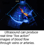
Ultrasound can produce a "live-action" moving image of functioning organs, and is useful in investigating blood flow through arteries and veins.
Ultrasound is useful for a wide variety of purposes
Because ultrasound produces accurate images of soft tissue, it's valuable as a tool for heart and vascular medicine to evaluate blood flow or possible clots or aneurysms in arteries and veins; and it provides detailed images of the size and function of the heart.
Ultrasound is also a good tool for evaluation of the size and function of other organs such as the gallbladder, liver, spleen and others. It can also be used to detect internal fluid, cysts, tumors or abscesses. Obstetric medicine uses ultrasound to provide detailed images of the fetus and uterus.
A safe and noninvasive diagnostic tool.
Because ultrasound uses only sound waves, it presents no risk of exposure to ionized radiation (such as X-rays). So it's very safe for pregnant women and unborn infants, and can be repeated as often as necessary.
Ultrasound is noninvasive, so it's almost always painless, and the equipment is less expensive, making it a very cost-effective diagnostic option.
For more information about radiology services at the main campus, please call (229) 312-1176.